GUEST EXPERT
ON THE BENEFITS OF CONCURRENT TMS-FMRI
In this article, Olivier Reynaud, coordinator of the Human Neuroscience Platform (HNP) and manager of the HNP Neuromodulation MRI facility, explains some benefits of the concurrent TMS-fMRI setup at Campus Biotech.
Multimodal imaging at Campus Biotech
Concurrent TMS-fMRI is a neuroimaging technique that combines the advantages of transcranial magnetic stimulation (TMS) and functional magnetic resonance imaging (fMRI) to study the brain. This technique offers several benefits over traditional TMS or fMRI alone, making it a valuable tool for advancing our understanding of brain function.
The concurrent TMS-fMRI setup is readily available to all users of the HNP.

The concurrent TMS-fMRI setup consists of an MRI-compatible TMS coil (white) delivering precise stimulation inside the MRI bore, and two dedicated MRI surface coils (blue) compatible with TMS and providing full brain coverage. Additional details on the TMS stimulator capabilities can be found HERE.
Advantages of the concurrent tms-fmri setup
Proof of target engagement
One of the major benefits of concurrent TMS-fMRI is its ability to provide proof of target engagement, or the ability to demonstrate that the TMS pulse is effectively stimulating the intended brain region. Traditional TMS is limited by the fact that it cannot provide direct evidence that the TMS pulse is reaching the intended brain region. fMRI, on the other hand, can be used to measure the brain’s response to TMS stimulation, providing proof of target engagement and allowing researchers to confirm that the TMS pulse is effectively stimulating the intended brain region.
Network connectivity
Another benefit of concurrent TMS-fMRI is its ability to study specific brain network connectivity. TMS allows researchers to study the connections between different brain areas, while fMRI provides information about the strength of these connections. By combining TMS and fMRI, researchers can study the functional connectivity of brain networks and disturb specific networks of interest, providing insight into how different brain regions interact and communicate with each other. This can be particularly useful for studying the organization and function of brain networks in both healthy individuals and individuals with neurological or psychiatric disorders.
Causality
In addition, concurrent TMS-fMRI also has the ability to establish causality, or the ability to demonstrate that a specific brain region or network is causally involved in a particular cognitive or behavioral process. By targeting a specific brain region with TMS, researchers can temporarily disrupt its function in a reversible manner, creating a virtual lesion in that region. fMRI can be used to measure the brain’s response to that TMS stimulation during a particular task, providing evidence of the causal involvement of the stimulated brain region in a particular process.
Support from the Human Neuroscience Platform
At Campus Biotech in Geneva, concurrent TMS-fMRI is available through the HNP MRI and Neuromodulation facilities. The HNP team provides full support for researchers interested in using this neuroimaging technique, including assistance with study design, implementation, and data analysis.
The HNP team at Campus Biotech has extensive experience with concurrent TMS-fMRI, and is equipped with state-of-the-art equipment and infrastructure to support this technique. Researchers can access a 3T MRI scanner and a TMS system that are specifically designed for concurrent TMS-fMRI, allowing for high-quality imaging and reliable results. The HNP team also provides training and support for researchers who are new to concurrent TMS-fMRI, ensuring that they have the knowledge and skills needed to successfully implement and analyze their experiments.
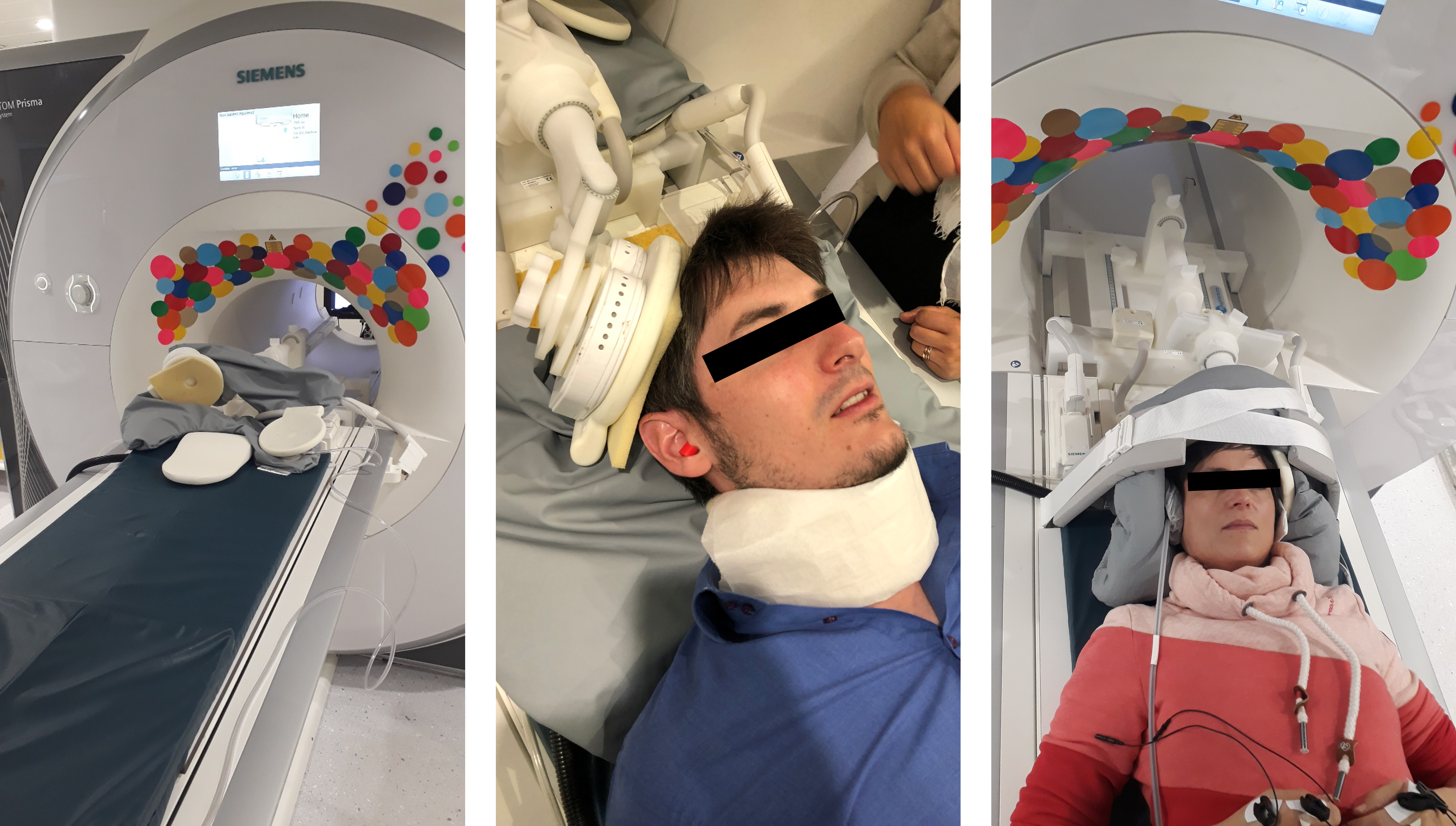
Examples of concurrent TMS-fMRI setups at Campus Biotech. From left to right: before, during and after volunteer installation.